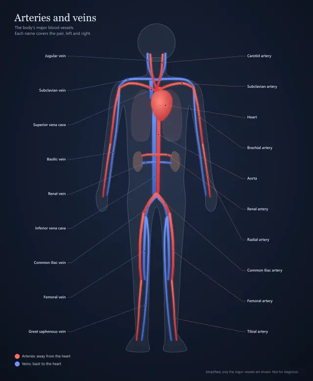
Superior vena cava
Brings oxygen-poor blood from the head, neck, arms and upper chest into the right atrium.
- Blood
- Oxygen-poor
- Flows into
- Right atrium
- Blood flow
- Step 1 of 12
A rich illustration under invisible regions: each part opens a panel, numbered along the blood's path.
The same content as the picture, as text: every part and what it says when you open it.
Brings oxygen-poor blood from the head, neck, arms and upper chest into the right atrium.
Brings oxygen-poor blood from the lower body into the right atrium. It is the largest vein in the body.
Collects oxygen-poor blood returning from the body and passes it down through the tricuspid valve.
Three leaflets between the right atrium and the right ventricle. It shuts as the ventricle contracts, so blood cannot flow back.
Pumps oxygen-poor blood out to the lungs. Its wall is thinner than the left ventricle's, because the lungs need much less pressure than the whole body.
Guards the way out of the right ventricle into the pulmonary artery, and closes so blood cannot slide back.
Carries blood from the right ventricle to both lungs, where it picks up oxygen. Unusually for an artery, the blood in it is oxygen-poor.
Bring oxygen-rich blood back from the lungs into the left atrium. There are usually four, two from each lung; the pair from the right lung runs behind the heart and is hidden here.
Collects oxygen-rich blood from the lungs and passes it down through the mitral valve.
Two leaflets between the left atrium and the left ventricle, which is why it is also called the bicuspid valve.
The heart's main pump. Its thick muscular wall drives oxygen-rich blood out through the aorta to the whole body.
Opens as the left ventricle contracts and closes behind each beat, so blood cannot return from the aorta.
The body's largest artery. It rises from the left ventricle, arches over the heart, and carries oxygen-rich blood on to every organ.
The muscular wall between the left and right sides, which keeps oxygen-rich and oxygen-poor blood apart.
Separates: Left and right sides
The heart muscle itself, which contracts with every beat. It is fed by its own coronary arteries, which run over its outer surface.
The heart's natural pacemaker, in the wall of the right atrium. Each heartbeat starts here as an electrical signal.
Holds the signal for a moment, so the atria finish emptying before the ventricles contract.
Carry the signal down the septum and out through the ventricle walls, so the ventricles squeeze from the bottom up.
Anatomy, science and history, the way a textbook would explain each part.